Mission
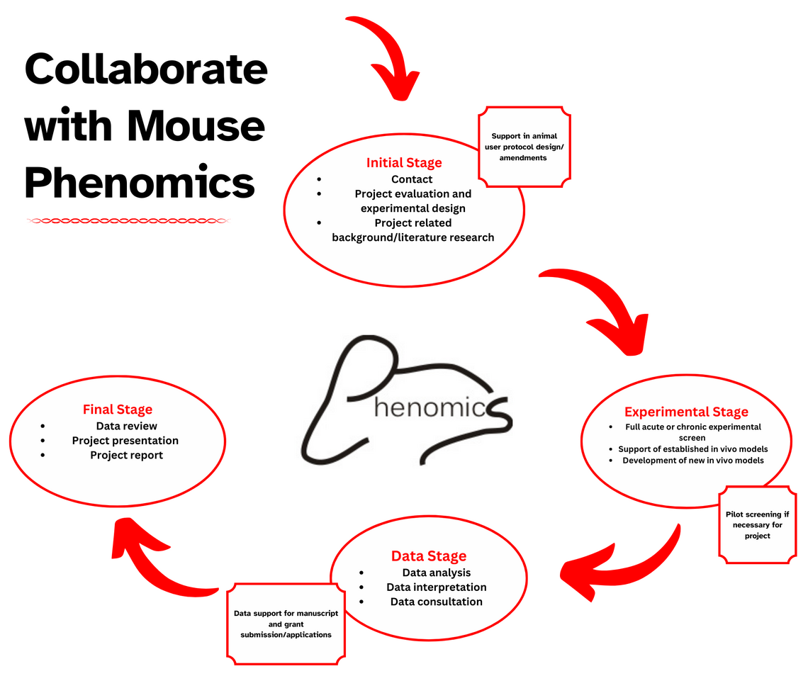
The mission of the Mouse Phenomics Resource Laboratory is to assist researchers within the institute and beyond as well as industry in promoting high quality, world-class research by providing support for their own driven in vivo models of interest and by generating additional novel data through the development of new in vivo screens and murine models in the field of acute and chronic inflammation and related chronic diseases.
Services
- Initiation of new Institute teams based on in vivo research; experimental & analytical support
- Dev't. of new in vivo screening techniques (acute & chronic disease models, non-invasive imaging techniques)
- Experimental design & data analysis for manuscript submissions & grant applications
- Dev't. of new mouse KO strains in collaboration with the CCCMG
- Experimental & analytical support re: mouse work for labs lacking in vivo experience
- Completion & further dev't. of ongoing experiments based on resource labs' in vivo experience
Key Areas of Research
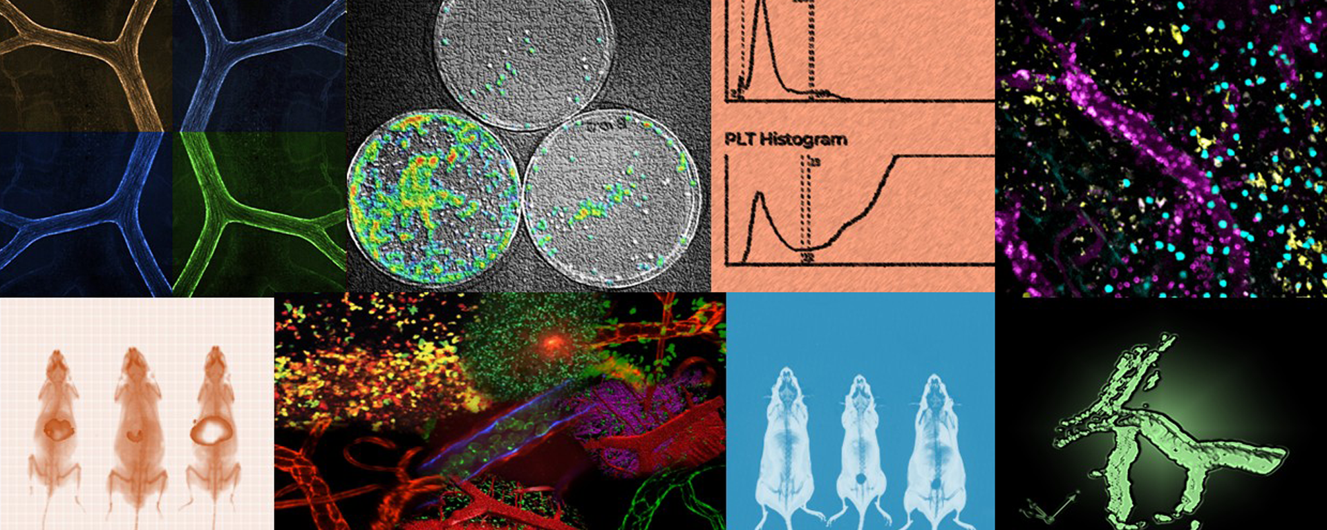
- Intravital microscopy of various organ systems/vascular beds
- acute and chronic non-invasive whole animal imaging screens
- Drug dissemination/location screens
- Acute and chronic infectious screens
- Acute and chronic inflammatory screens
- Murine models of human chronic diseases
- Metabolic screens
- Hematology analysis
Select Publications
- Tan X. et al., Mol Microbiol. 2021 (Real time tracking of borrelia infection via IVM)
- Ryes J.L. et al., FASEB J 2019 (macrophage assoc. colitis incl. whole body imaging)
- Zeng Z. et al., Nat Immunol. 2018 (Ab assoc. protection from bact. infection incl. whole body imaging)
- Andonegui G. et al., JCI Insight 2018 (sepsis assoc. brain damage incl. whole body imaging)
- Kim J.H. et al., Nat Immunol. 2018 (lung inflammation & arthritis model)
- Ramachandran R. et al., Mol Pharmacol. 2017 (intravital drug screen)
- Leung G. et al., Mol. Med. 2015 (GI inflammation, incl. whole body imaging)
- Lopes F. et al., Arthritis Rheumatol. 2015 (arthritis model)
Contacts
Dr. Björn Petri
Scientific Director: Mouse Phenomics Resource Laboratory, Calvin, Phoebe and Joan Snyder Institute for Chronic Diseases Research Associate Professor Dept. Microbiology, Immunology & Infectious Diseases Cumming School of Medicine, University of Calgary
HSC 2868 (lab): 403.210.6538
HSC 2825 (office): 403.220.4562
Email: bpetri@ucalgary.ca
Getting Started with Mouse Phenomics

Core Personnel
Bjöern Petri, PhD, MSc
Scientific Director, Research Associate Prof
Dr. Bjӧern Petri is the Scientific Director of the Snyder Institute Mouse Phenomics Resource Laboratory since January 2011. Dr. Petri obtained his PhD (Dr. rer. nat.) in Immunology/Cell Biology and Genetics from the Westfalian Wilhelms-University and the Max-Planck-Institute for Molecular Biomedicine in Münster, Germany. Before he started his studies in the field of Immunology Dr. Petri obtained his MSc (Diploma) in Zoology and Physiology at the Max-Planck-Institute for Heart and Lung Research in Bad Nauheim and the Philipps-University in Marburg, Germany. He can look back at over 20 years of in vivo experience working with rodent models in the field of physiology and immunology.
Between 2007 and 2010 he worked as a Postdoctoral Fellow (AHFMR/AIHS) in the Snyder Institute for Chronic Diseases where he continued to strengthen his expertise in the field of Immunology and its related research models and imaging techniques.
Specialization / Applications
Acute inflammatory screens in WT and KO mice:
- Bacterial skin infection.
- Leukocyte recruitment screens in different vascular beds under physiological and infectious/inflammatory conditions.
- Permeability models under physiological and infectious/inflammatory conditions in different vascular beds.
Murine models of human chronic diseases in WT and KO mice:
- Chronic bacterial infections/sepsis.
- Serum transfer induced arthritis (K/BxN model).
- TNBS/DSS models of colitis and AOM/DSS models of colitis associated cancer.
- DTH model.
- BMT/chimeric screens.
- Access to additional models within the Snyder Institute as well as metabolic screening tools.
Intravital and non-invasive whole body imaging techniques as screening tools.
- Intravital imaging of different murine organs/vascular beds using spinning-disk confocal and multiphoton microscopy under physiological and pathological/disease conditions (in collaboration with the Live Cell Imaging Resource Laboratory).
- Development/testing of new imaging reagents/probes using non-invasive whole body imaging (Spectral Instruments Imaging AMI HTX).
- Non-invasive whole body imaging screens under physiological and pathological/disease conditions.


